Times Health Critical Care Survey - NU Hospitals ranked 1st for City , 2nd for Regional and 3rd for National in the field of Urology. Times Health Critical Care Survey - NU Hospitals ranked 6th for City in the field of Nephrology

Kidney Cancer

Kidney cancer, also called renal cancer, is an ailment in which kidney cells become malignant (cancerous) and grow out of control, forming a tumor. Almost all kidney cancers first appear in the lining of tiny tubes in the kidney. This type of kidney cancer is called renal cell carcinoma.
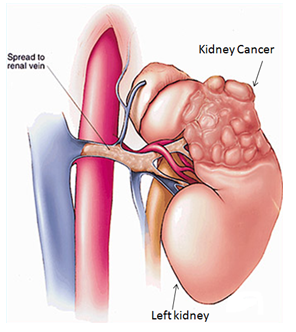
What is the role of kidneys in our body?

The kidneys are a matched pair of vital organs located underneath your liver and stomach and near your backbone on either side. They help remove wastes from the body by making urine. They do this by filtering urea, salt and other substances from the blood as it flows through the kidneys. The kidneys serve also as glands that manufacture and secrete a variety of hormones.

What are the types of kidney tumors?

A kidney tumor is an abnormal growth within the kidney. Tumors may be benign or malignant.

Benign tumors (non-cancerous) remain localized and usually do not spread or threaten one’s life. The most common kidney lesion seen on ultrasound is a fluid-filled area called renal cyst. Simple cysts are benign and have a typical appearance on ultrasound studies and usually don’t require any treatment.

Malignant tumors are cancers. They can invade and destroy nearby tissues and organs or spread to other parts of the body by way of the bloodstream or lymphatic system. More than 80% of solid renal lesions are cancerous.

How common are malignant renal tumors?

It accounts for 2% to 3% of all adult malignant neo­plasms. The typical presentation is in the sixth and seventh decades of life. Male-to-female pre­dominance of 3:2.

What are the risk factors associated with kidney cancer?

  • Smoking
  • Hypertension
  • Obesity
  • Family history of kidney cancer
  • Chronic kidney failure and/or dialysis
  • Diet with high fat
  • Certain genetic diseases involving kidneys (Von Hippel-Lindau syndrome, tuberous sclerosis)

Are there any symptoms related to kidney cancer?

Today up to 50 % of new kidney tumors are detected incidentally during routine health check or during evaluation for some unrelated problem. Symptoms of kidney cancer are blood in the urine, lump or mass in the abdomen and pain in the side. If tumors spread beyond the kidney, the patient may present with fatigue, loss of appetite, weight loss and anemia.

How is kidney cancer diagnosed?

Diagnosis begins with a physical examination by your doctor. Diagnosis is usually first made based on Ultrasound study and later confirmed by Computed Tomography of the abdomen. Your doctor will perform other tests that will tell him whether cancer has spread beyond the kidney or not. These include Chest X-ray, radioisotope bone scan (if the patient has bone pain, recent bone fractures or certain abnormalities of blood tests).

What are the different stages?

Based on all investigations and findings, your doctor will clinically stage the tumor. The factors considered for staging are the size of the tumor (less than 4 cm, 4-7 cm, 7-10 cm or more than 10 cm), if it is confined to the kidney, spread to the renal veins or to surrounding tissues and organs or distant organs.

What are the treatment options?

Your medical history, general health, the size, location of cancer and many other factors are considered in determining the treatment needed.

Surgery: Is the standard treatment for most cases of adult kidney cancers that have not spread to distant areas of the body. Based on location, size your doctor will decide if only a part of the kidney (partial nephrectomy) or entire kidney needs to be removed (radical nephrectomy). This operation can be done by traditional open technique or by keyhole incisions (laparoscopic) and this will be decided based on CT scan and technical accessibility of the tumor. The goal of partial nephrectomy is to remove the entire tumor while preserving as much normal kidney tissue as possible.

Immunotherapy: This group of drugs stimulates your immune system to attack cancer. These drugs are used in patients where the tumor has spread beyond the kidney to other organs. But it has shown some decrease in tumor size in only 20% of patients. These drugs are given intravenously either before or after nephrectomy.

Targeted Agents: These drugs cause the vessels supplying the tumor to regress. It can slow the progress of kidney cancer and allow patients to live longer. These drugs are taken orally but they can be associated with side effects including fatigue, hypertension and skin problems. But most patients are able to tolerate these drugs fairly well and appear to benefit from them. Like immunotherapy, these drugs can be used either before or after nephrectomy. The other drawback is these drugs are expensive at present.

When will I know the final report?

The kidney/tumor that is removed will be sent for pathological examination and report should be available in 1 week time. With this report, the doctor will pathologically stage your disease and advice accordingly.

Is there any need for follow-up?

Yes. To look for any local or distant recurrence and care of the remaining kidney. The frequency of follow up will be based on stage and tumor characteristics.

Will my kidney function deteriorate after this surgery?

When one kidney or part of a kidney is removed, the remaining functional kidney tissue usually works sufficiently to avoid problems, if there is a normal kidney on the other side. In fact, people can live a normal life with only one functioning kidney. Routine follow ups of kidney function is recommended. Some medical conditions, such as high blood pressure (hypertension), diabetes, high cholesterol and obesity, have the potential to cause deterioration of kidney function. Treatment of these conditions may prevent kidney damage. Therefore, it is important that you comply with Nephrologist supervised treatment of these conditions.

Urinary Bladder Cancer

The inside, or inner lining, of the bladder is called Urothelium. Next to this is a layer of loose connective tissue called the Lamina Propria. Deeper to this is the bladder muscle and the outer most layer is fat. When found and treated in the early stages, bladder cancers are not likely to be life threatening.

What is the Urinary Bladder?

The bladder is that part of the urinary tract that stores urine until the person is ready to pass urine. Urine is produced by the kidneys. Urine travels from the kidney down a narrow tube, the ureter and is stored in balloon-like muscular organ, the bladder. Urine is discharged through the urethra during urination.

What is Urinary Bladder Cancer?

Occasionally, if these urothelial cells start to multiply uncontrollably a new growth or tumour starts.

When found and treated in the early stages, bladder cancers are not likely to be life threatening. In addition, treatment of most of these tumors does not require removal of the bladder. Prompt medical attention and regular checkups are necessary to treat bladder tumors and to watch for new growths.

How common is Urinary Bladder Cancer?

It is the 9th most common cancer worldwide. The median age of bladder cancer diagnosis is 70 years.

Who is at risk of getting bladder cancer?

Bladder cancer is unusual in people under 40 years of age. Men are affected 3 to 4 times more often than women and cigarette smokers have an increased risk of developing bladder cancer. Exposure to certain industrial chemicals (paints and aromatic amines) in the workplace also has been associated with an increased risk of developing bladder cancer.

What are the symptoms of Urinary Bladder Cancer?

The earliest clue of bladder tumour is the presence of blood in your urine. Blood in the urine is usually not accompanied by pain, for those with bladder cancer. Sometimes you may not see the blood in urine and it can be seen only under a microscope. People with kidney stones or urinary tract infections and men with enlarged prostate glands may also have blood in their urine. Blood in urine is a potential warning sign of cancer and should not be ignored. Other symptoms include frequent urination or burning urination in the absence of urinary infection.

How is Urinary Bladder Cancer diagnosed?

Your doctor will perform further tests after physical examination.  These tests may include urine analysis, urine cytology (to look for cancer cells that may have been shed into the urine from the bladder lining), ultrasound study of the abdomen, contrast CT (computed tomography) of the abdomen. CT will tell if there are other similar lesions in the kidneys and ureter. In addition to the bladder details, CT also tells us involvement of surrounding organs. In most of the cases cystoscopic examination under anesthesia (medication that puts you to sleep) is mandatory. All of the tests above may be performed by an urologist without requiring you to stay overnight in a hospital.

Cystoscopy

In cystoscopy, an endoscope (cystoscope) is inserted gently into the urethra and passed into the bladder to directly view the inside of the bladder lining. This is done under I.V. sedation. If any tumor is found you will be rescheduled for resection biopsy (TUR-BT i.e., Transurethral resection of bladder tumor) under spinal or general anesthesia. Based on the biopsy report, further treatment plan would be discussed with you.

On what basis is the Urinary Bladder Cancer treatment planned?

The treatment for bladder cancer depends on how deeply the tumour has grown into the bladder wall. The growth may be superficial, i.e., it is confined to urothelium and lamina propria or deep muscle invasive, if it involves the bladder muscle. Also the size, location and number of tumors are noted. During resection both the superficial and deep tissues are sent separately for biopsy testing. The biopsy report also gives the grade of tumor (either low or high grade). With all these clinical details, along with age, co-morbidities, performance score your doctor will decide on further treatment.

Will further treatment be needed?

If the doctor believes that you are likely to develop new cancer (recurrence), you may be advised to undergo additional treatments like having medications instilled into the bladder (refer to BCG brochure).

Some bladder cancers are muscle invasive. In such cases, the urologist may recommend that the bladder be completely removed and urine will be diverted by one of the several methods. This operation is called a cystectomy. The doctor may also recommend additional treatment with radiation and chemotherapy drugs. Regular follow up is required because bladder cancers often recur, especially within the first year or two after discovery of the first cancer. Because tumours can recur, it is important for the urologist to do Cystoscopy and cytology regularly.

What can be done to prevent the recurrence of bladder cancer?

Smoking cessation, increased fluid intake, and a low-fat diet may all reduce the risk of recurrence. Second, is by regular follow-up visits and cystoscopy as advised by your doctor. Third, is by medications instilled into the bladder.

Is there any need for follow-up?

Yes. To look for any local or distant recurrence and care of the remaining kidney. The frequency of follow up will be based on stage and tumor characteristics.

Public Notice: NU Hospitals would like to inform the general public that NU Hospitals practices all organ transplants in accordance with The Transplantation of Human Organs Act 1994. NU Hospitals does not buy or sell any organ and seriously condemn this act. NU Hospitals do not by any nature seek your personal information such as name, telephone, address or banking details for any purpose.